

A photomicrograph is a technical document that can be of great significance to science. A one is also an object of beauty, open to several levels of appreciation. And once a year, Nikon celebrates the best of the bunch.
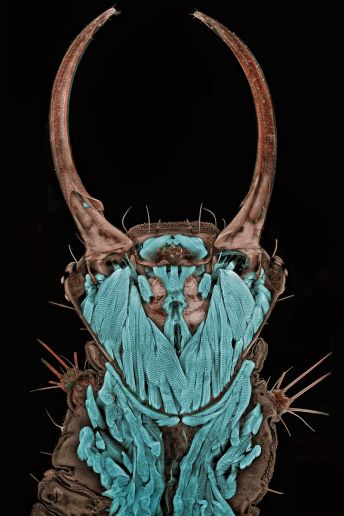
1st Place -- Dr. Igor Siwanowicz
Portrait of a green lacewing larva
Technique: Confocal, magnified 20X
Source: Dr. Igor Siwanowicz/Max Planck Institute of Neurobiology

2nd Place -- Dr. Donna Stolz
Blade of grass
Technique: Confocal stack reconstruction, autofluorescence, magnified 200X
Source: Dr. Donna Stolz/University of Pittsburgh

3rd Place -- Frank Fox
Melosira monoliformis (living specimen)
Technique: Differential interference contrast, magnified 320X
Source: Frank Fox/Fachochschule Trier

4th Place -- Dr. Robin Young
Liverwort
Technique: Live mount, confocal microscopy, magnified 20X
Source: Dr. Robin Young/University of British Columbia

5th Place -- Alfred Pasieka
3D reconstruction of a microchip
Technique: Incident light, Normarski interference contrast, magnified 500X
Source: Alfred Pasieka

6th Place -- Dennis Callahan
Cracked solar cell films
Technique: Brightfield, magnified 50X
Source: Dennis Callahan/California Institute of Technology

7th Place -- Gabriel Luna
Nerve fibers from the retina of a mouse
Technique: Laser confocal scanning, magnified 40X
Source: Gabriel Luna/UC Santa Barbara, Neuroscience Research Institute

8th Place -- Dr. Bernardo Cesare
Coarse-grained rocks bearing graphite
Technique: Polarized light, magnified 2.5X
Source: Dr. Bernardo Cesare/Department of Geosciences

9th Place -- Dr. Jan Michels
The underbelly of a marine copepod
Technique: Confocal, autofluorescence and congo red fluorescence, magnified 10X
Source: Dr. Jan Michels/Christian-Albrechts-Unversitat zu Kiel

10th Place -- Joan Rohl
Freshwater water flea
Technique: Differential interference contrast, magnified 100X
Source: Joan Rohl/Institute for Biochemistry and Biology

11th Place -- Dr. Jan Michels
Front view of an ant head
Technique: Confocal, autofluorescence, magnified 10X
Source: Dr. Jan Michels/Christian-Albrechts-Universitat zu Kiel

12th Place -- Thomas Deerinck
"Immortal" cancer (HeLa) cells
Technique: 2-Photon fluorescence, magnified 300X
Source: Thomas Deerinck/National Center for Microscopy and Imaging ResearchV

13th Place -- Dr. Stephen S. Nagy
A cross-section of a curare vine
Technique: Brightfield, digitally inverted, magnified 45X
Source: Dr. Stephen S. Nagy/Montana Diatoms

14th Place -- Yanping Wang
Various grains of sand
Technique: Reflected light, magnified 4X
Source: Yanping Wang/Beijing Planetarium

15th Place-- James H. Nicholson
A live specimen of lobe coral, its tissue pigmented with red fluorescence
Technique: Epiflurescence with triple band (U/B/G) excitation, magnified 12X
Source: JAmes H. Nicholson/Coral Culture and Collborative Research Facility


